Introduction
Dark field microscopes are essential tools in the realm of microscopy, providing unique imaging capabilities that are indispensable for various scientific and medical applications. Unlike traditional light microscopes, dark field microscopes enhance contrast in specimens that are otherwise transparent and difficult to observe. This article delves into the principles, applications, advantages, and limitations of dark field microscopes, offering a comprehensive guide to understanding this sophisticated optical instrument.

Understanding Dark Field Microscopy
What Is Dark Field Microscopy?
Dark field microscopy is a technique that illuminates a specimen with light that is not directly visible through the microscope objective. Instead, it relies on scattered light to produce a contrast-rich image against a dark background. This method is particularly useful for visualizing specimens that are too small or too transparent to be seen with conventional light microscopy.
How Dark Field Microscopes Work
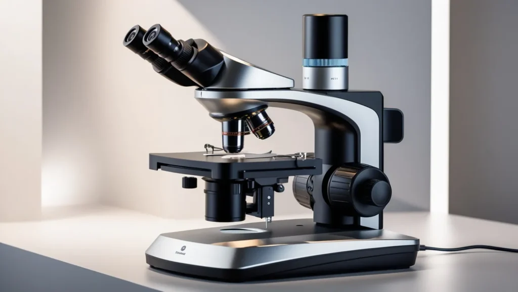
Dark field microscopes operate by using a special condenser that directs light at an angle onto the specimen. The key components include:
- Dark Field Condenser: This is an optical element that blocks direct light from entering the objective lens. It allows only scattered light from the specimen to pass through.
- Objective Lens: Captures the scattered light that has interacted with the specimen, forming an image.
- Illumination System: Usually includes a light source that illuminates the specimen at an oblique angle.
The Principle of Dark Field Imaging
The fundamental principle of dark field imaging lies in its ability to enhance contrast by exploiting light scattering. When light hits a specimen, it may scatter in different directions depending on the specimen’s properties. The dark field microscope captures only the scattered light, leaving the non-scattered, direct light out of the image. This creates a high-contrast image where the specimen appears bright against a dark background.
Key Components of Dark Field Microscopes
Dark Field Condenser
The dark field condenser is the heart of a dark field microscope. It is designed to focus light onto the specimen at a very oblique angle. This design ensures that the light entering the objective lens is only the light scattered by the specimen, rather than the direct light that would otherwise create a bright field image.
Objective Lens
The objective lens in a dark field microscope is critical for capturing the scattered light. High numerical aperture (NA) objectives are often used to collect as much scattered light as possible, enhancing image resolution and contrast.
Illumination System
The illumination system in dark field microscopes typically includes a light source, such as a halogen or LED lamp, which is focused through the condenser. The choice of light source can affect the quality and contrast of the images produced.
Sample Stage
The sample stage holds the specimen and allows for precise movement and positioning. In dark field microscopy, the sample stage needs to be stable and adjustable to ensure accurate alignment and observation.
Applications of Dark Field Microscopes
Biological Research
In biological research, dark field microscopes are used to study live cells, bacteria, and other microorganisms. Its ability to enhance contrast allows researchers to observe fine details and structures without the need for staining, which can alter or damage the specimen.
Medical Diagnostics
Dark field microscopes are employed in medical diagnostics to detect and analyze various pathogens, including spirochetes and other bacteria. It is particularly useful for diagnosing diseases like syphilis, where dark field microscopy can reveal the characteristic spiral-shaped bacteria.
Material Science
In material science, dark field microscopes help analyze the structural properties of materials, such as metals, crystals, and polymers. It is used to study surface features, grain boundaries, and other microscopic details that are crucial for understanding material properties.
Forensic Science
Forensic scientists use dark field microscopes to examine traces of evidence, such as fibers, hairs, and particles. The high contrast and detailed imaging capabilities of dark field microscopy make it an invaluable tool in forensic investigations.
Advantages of Dark Field Microscopes
Enhanced Contrast
One of the primary advantages of dark field microscopes is their ability to provide high contrast images of transparent and small specimens. This enhanced contrast allows for better visualization of fine details that are often missed with traditional microscopy techniques.
No Staining Required
Dark field microscopy does not require staining or chemical treatments, which preserves the natural state of the specimen. This is particularly important for live cell imaging and sensitive biological samples.
Visualization of Small Structures
The technique is excellent for visualizing small structures, such as microorganisms and nanoparticles, which may be challenging to observe with standard light microscopy.
Improved Resolution
By capturing scattered light, dark field microscopes can offer improved resolution compared to bright field microscopy. This allows for the observation of finer details and structures within the specimen.
Limitations of Dark Field Microscopes
Limited Depth of Field
Dark field microscopy often has a limited depth of field, which can make it challenging to focus on thicker specimens. This limitation can be mitigated by using techniques such as focusing adjustments or alternative imaging methods.
High Light Intensity
The high-intensity illumination required for dark field microscopy can sometimes lead to specimen damage or photobleaching. Careful control of light intensity and exposure time is essential to prevent such issues.
Artifact Presence
Dark field microscopes may produce artifacts, such as stray light or reflections, which can interfere with image quality. Proper calibration and adjustment of the microscope can help minimize these artifacts.
Cost and Complexity
Dark field microscopes can be more expensive and complex than standard light microscopes. The specialized components and alignment requirements contribute to the overall cost and complexity of the system.
How to Set Up a Dark Field Microscope
Assembling the Microscope
- Install the Light Source: Place the light source in its designated position and ensure it is properly aligned with the condenser.
- Attach the Dark Field Condenser: Install the dark field condenser into the microscope, adjusting it to the correct position for optimal illumination.
- Insert the Objective Lens: Choose a high NA objective lens and insert it into the microscope’s objective turret.
- Prepare the Sample Stage: Place the specimen on the sample stage and adjust its position for precise alignment with the condenser.
Adjusting the Illumination
- Set the Light Intensity: Adjust the light intensity to achieve the desired contrast and prevent overexposure of the specimen.
- Align the Condenser: Ensure the dark field condenser is correctly aligned with the specimen to achieve optimal illumination.
Focusing and Imaging
- Focus the Microscope: Use the coarse and fine focus controls to bring the specimen into clear view.
- Capture Images: Once focused, capture images using a camera or digital imaging system if available.
Conclusion
Dark field microscopes represent a powerful and versatile tool in microscopy, offering unique advantages for visualizing transparent and small specimens. By understanding the principles, components, and applications of dark field microscopes, researchers and scientists can leverage this technique to gain deeper insights into various fields, including biology, medicine, material science, and forensic science. Despite its limitations, the enhanced contrast and resolution provided by dark field microscopy make it an invaluable asset in the quest for detailed and accurate observations.
FAQs
1. What types of specimens are best suited for dark field microscopy?
Dark field microscopes are ideal for observing transparent specimens, such as live cells, bacteria, and small particles, which are difficult to visualize with traditional light microscopy.
2. How does dark field microscopy compare to bright field microscopy?
Dark field microscopy offers higher contrast and resolution for transparent specimens, while bright field microscopy provides a more straightforward view of stained or colored specimens.
3. Can dark field microscopy be used for quantitative analysis?
While dark field microscopy excels in visualizing details and structures, it is generally less suited for quantitative analysis compared to other techniques like fluorescence microscopy.
4. Are there any specific maintenance requirements for dark field microscopes?
Regular maintenance includes cleaning optical components, calibrating the alignment of the condenser and light source, and checking for any mechanical issues.
5. What are common applications of dark field microscopy in clinical diagnostics?
Dark field microscopes are commonly used in clinical diagnostics to detect pathogens such as spirochetes and other bacteria, which are crucial for diagnosing diseases like syphilis.

