Introduction
In the realm of microscopy, the ability to accurately capture images of microscopic organisms and structures is pivotal. Whether you’re delving into the study of bacteria or observing intricate crystals in water, the level of zoom—or magnification—required is a crucial factor that determines the clarity and detail of your images. This comprehensive guide explores the specific magnification levels needed to photograph bacteria and crystals effectively, providing in-depth insights into microscope types, imaging techniques, and practical considerations.

Understanding Microscope Magnification
The Basics of Magnification
Magnification is the process of enlarging the appearance of an object through optical means. In microscopy, this involves the use of lenses to increase the apparent size of tiny specimens. The effectiveness of magnification is measured in terms of magnification power, which is the degree to which the microscope enlarges the image of the specimen.
Types of Microscopes
Light Microscopes
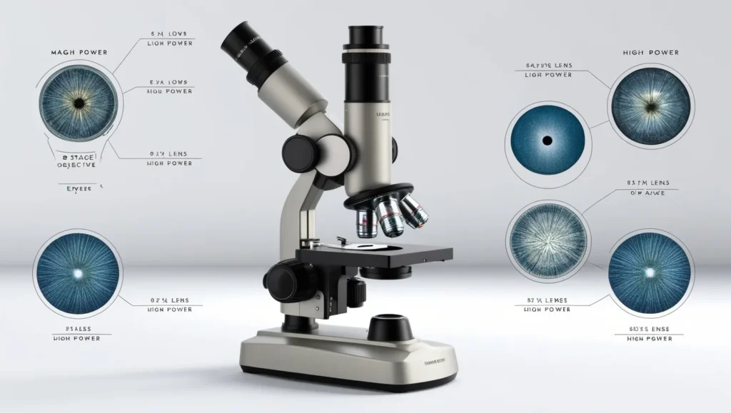
Light microscopes use visible light and lenses to magnify specimens. They are commonly used for observing live bacteria and crystals. Their magnification typically ranges from 40x to 1000x, depending on the objective lenses used.
Electron Microscopes
Electron microscopes use electron beams instead of light, allowing for much higher magnifications, from 10,000x to 2,000,000x. These are essential for detailed imaging of bacteria and crystals at the molecular level but are not typically used for standard laboratory photography.
Magnification Requirements for Photographing Bacteria
Choosing the Right Objective Lens
To photograph bacteria, you need to achieve sufficient magnification to resolve their detailed structures. The 100x oil immersion lens is often the optimal choice, as it provides a high magnification power and a large numerical aperture, which enhances resolution and contrast.
Resolution and Image Clarity
Resolution is the ability of the microscope to distinguish between two points that are close together. For photographing bacteria, a resolution of 0.2 micrometers is typically required to view individual cells and their structures clearly. This is achievable with high-quality light microscopes using appropriate magnification and objective lenses.
Photographing Crystals in Water
Understanding Crystal Structures
Crystals in water can vary greatly in size and complexity. Observing these crystals requires precise magnification to reveal their intricate structures and facets. Polarizing light microscopes are particularly useful for studying crystals as they can enhance contrast and reveal details not visible under regular light.
Optimal Magnification Levels
For photographing crystals, a magnification range of 200x to 400x is generally effective. This level allows for a clear view of the crystal’s geometry and internal features. Higher magnifications may be necessary for more detailed studies, especially for larger or more complex crystals.
Practical Considerations for Microscopic Photography
Sample Preparation
Proper sample preparation is crucial for achieving high-quality images. For bacteria, samples should be stained to enhance contrast. Crystals in water should be carefully positioned and, if necessary, isolated to prevent overlapping and distortion.
Lighting and Contrast
Lighting plays a significant role in microscopic photography. For bacteria, bright-field illumination is commonly used, while polarized light is essential for crystals to reveal their unique properties.
Camera and Imaging Techniques
The choice of camera and imaging technique also affects the quality of your photographs. Digital microscopes equipped with high-resolution cameras offer the advantage of direct imaging, while traditional microscopes may require camera adapters and specialized settings for optimal results.
Choosing the Right Equipment
Microscope Types and Their Applications
Bright-Field Microscopes
Ideal for general observation and photography of bacteria and simple crystal structures. They provide clear images with standard contrast.
Phase-Contrast Microscopes
Useful for observing live bacteria without staining, preserving their natural state and providing better contrast.
Polarizing Microscopes
Essential for studying crystals due to their ability to enhance and reveal the details of crystal structures through polarized light.
Investing in High-Quality Lenses
The quality of your objective lenses impacts the final image quality. Investing in high-quality lenses with excellent resolution and numerical aperture will significantly improve the clarity of your photographs.
Troubleshooting Common Issues
Blurry Images
Blurriness can result from improper focusing, poor lighting, or low-quality lenses. Ensure that the microscope is correctly focused, and consider adjusting the lighting or upgrading the lens if necessary.
Low Contrast
Low contrast can be addressed by adjusting the lighting conditions or using staining techniques for bacteria. For crystals, ensuring proper polarization can enhance contrast and reveal intricate details.
Overlapping Specimens
When photographing crystals, overlapping specimens can distort images. Use techniques to separate crystals or adjust the positioning to minimize overlap.
Conclusion
In summary, photographing bacteria and crystals under a microscope requires careful consideration of magnification levels and equipment. For bacteria, a magnification of 100x with oil immersion lenses typically provides the necessary detail, while crystals generally require 200x to 400x magnification for clear imaging. By choosing the right microscope, lenses, and imaging techniques, you can capture high-quality photographs that reveal the fascinating details of these microscopic entities.

FAQs
1. What is the best type of microscope for photographing bacteria?
The bright-field microscope with a 100x oil immersion lens is often the best choice for photographing bacteria due to its high magnification and resolution.
2. Can I use a standard camera with a microscope for photography?
Yes, you can use a standard camera with a microscope camera adapter. However, digital microscopes with built-in cameras offer better image quality and convenience.
3. How do I prepare samples of bacteria for microscopic photography?
Bacteria samples should be stained to enhance contrast and visibility. Use appropriate staining techniques based on the bacterial species and desired level of detail.
4. What is the role of polarized light in crystal photography?
Polarized light helps enhance contrast and reveal detailed structures of crystals that are not visible with regular light, making it essential for accurate crystal imaging.
5. How can I improve the contrast of my microscopic images?
Improving contrast can be achieved by adjusting lighting conditions, using staining techniques for bacteria, or utilizing polarized light for crystals. Ensure proper alignment and calibration of the microscope for optimal results.

